by
by Introduction to External Ventricular External Ventricular Drain Industry
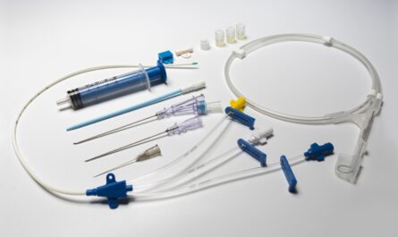
An external ventricular drain (EVD) is a drainage catheter that is placed in a ventricle of the brain to drain excess cerebrospinal fluid (CSF) from the brain and relieve elevated intracranial pressure. EVDs are often placed when hydrocephalus, traumatic brain injury, or other conditions cause a buildup of CSF in the brain ventricles that needs to be drained externally. They provide a direct method to sample CSF, monitor intracranial pressure, and drain fluid from the ventricles.
Indications for External Ventricular Drain Industry
Some common indications for EVD placement include:
– Hydrocephalus: Global External Ventricular Drain are often the initial treatment for hydrocephalus, whether it is communicating or obstructive in nature, to drain CSF and relieve elevated intracranial pressure.
– Intracerebral hemorrhage: An EVD may be placed after an intracerebral hemorrhage, such as a hemorrhagic stroke, to drain CSF and blood from around the brain. This can help prevent compression of brain tissue.
– Traumatic brain injury: In patients with severe traumatic brain injuries, an EVD may be placed to monitor and drain CSF, allowing the brain room to swell without increasing intracranial pressure.
– Infection/inflammation: Conditions causing infection or inflammation within the brain like meningitis or encephalitis may require an EVD to drain CSF and reduce pressure.
– Post-neurosurgery: An EVD is commonly placed after brain tumor resection or other neurosurgery to drain CSF during healing and prevent fluid buildup.
EVD Placement Procedure
The EVD placement procedure is typically done in the neuro ICU or operating room. Patients receive sedation or general anesthesia. The neurosurgeon then makes a small incision and burr hole in the scalp and uses imaging guidance like ultrasound or fluoroscopy to navigate the catheter through the brain tissue into one of the lateral ventricles. They then secure the catheter to the scalp. The distal end of the catheter is connected to an external drainage and monitoring system.
The system precisely measures CSF outflow and records intracranial pressure readings. Typically the goal is to keep pressure under 15-20 mmHg. The drainage system includes tubing and a collection chamber to catch and measure CSF output. Samples can also be taken from the catheter for analysis. The catheter site is cleaned and dressed until removal.
Nursing Care with External Ventricular Drain
Nursing care is crucial for EVD management to prevent complications and optimize patient outcomes. Key aspects of EVD nursing care include:
– Strict sterile technique is used for all connections and accessing the drainage system to prevent infections. Dressings are checked regularly.
– Monitoring intake and output of CSF closely. Target drainage rates are maintained based on the condition being treated.
– Checking intracranial pressure readings every 1-4 hours and notifying the neurosurgeon of any significant changes.
– Inspecting the catheter exit site for signs of drainage, redness or swelling which could indicate infection or CSF leak.
– Testing CSF samples as ordered to check for signs of infection or other abnormalities.
– Keeping the patient in a flat, supine position and avoiding movements that cause increases in intracranial pressure like extreme flexion, extension or coughing without support.
– Adjusting the height of the drainage system appropriately based on the patient’s position to regulate ICP.
– Watching for signs of over or under drainage like headache, seizures or altered mental status and reporting these promptly.
– Providing comfort measures like repositioning, pain medication and anti-nausea treatment as needed.
Potential Complications of External Drainage
While EVD treatment has many benefits, some potential complications can occur:
– Infection of the drain site or ventricles is a serious risk. Strict sterile technique during EVD access and care decreases this risk significantly.
– Hemorrhage can occasionally happen during catheter placement risks injury to vessels or brain tissue.
– Malfunction of the catheter like clogging, kinking or breakage may prevent proper CSF drainage and pressure monitoring.
– Over or under drainage of CSF can cause problems like subdural fluid collections, brain shift issues or rebound hyperacute pressure changes.
– Positioning the patient improperly carries a risk of increasing ICP during bending or coughing without support.
In Summary, careful nursing assessment and management play a key role in promptly identifying any issues so they can be addressed. With optimal care, EVDs provide an effective method for temporary CSF drainage and ICP monitoring in many neurological patients.
*Note:
1.Source: Coherent Market Insights, Public sources, Desk research
2.We have leveraged AI tools to mine information and compile it